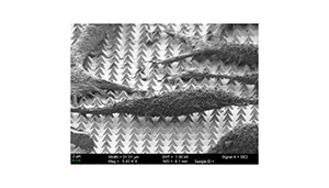
Researchers from the Harvard University’s John A. Paulson School of Engineering and Applied Sciences have developed a method to deliver molecules into cells using gold microstructures. The team published their work in ACS Nano.
Previous work has been done to deliver drugs or DNA into cells by tricking or forcing open the cell membrane, but the Harvard team says those methods are limited in the type of cargo they can carry.
“Being able to effectively deliver large and diverse cargos directly into cells will transform biomedical research,” 1st author Nabiha Saklayen said in prepared remarks. “However, no current single delivery system can do all the things you need to do at once. Intracellular delivery systems need to be highly efficient, scalable, and cost effective while at the same time able to carry diverse cargo and deliver it to specific cells on a surface without damage. It’s a really big challenge.”
The team used template-stripping as a fabrication method to make surfaces with 10 million, pyramid-shaped microstructures. Previously, they’ve demonstrated that these structures are good at focusing laser energy into electromagnetic hot-spots.
“The beautiful thing about this fabrication process is how simple it is,” coauthor Marinna Madrid said. “Template-stripping allows you to reuse silicon templates indefinitely. It takes less than a minute to make each substrate, and each substrate comes out perfectly uniform. That doesn’t happen very often in nanofabrication.”
After culturing the pyramids with HeLa cancer cells and surrounding them with a solution containing molecular cargo, the team used nanosecond laser pulses to heat the pyramids. They did so until the hot-spots at the tips of the pyramids reached 300 degrees Celsius, causing bubbles to form at the tip of each structure. The bubbles gently pushed into the cell membrane, creating pores and allowing the molecules to enter the cell, the team reported.
“We found that if we made these pores very quickly, the cells would heal themselves and we could keep them alive, healthy and dividing for many days,” Saklayen said.
Each cancer cell sat above 50 pyramids, so the team made about 50 pores in each cell. The team added that they could control the size of the bubbles by manipulating the laser parameters and could even control which side of the cell the cargo would penetrate.
The team said they plan to test the method on different cell types, such as blood cells, stem cells and T cells.
“This work is really exciting because there are so many different parameters we could optimize to allow this method to work across many different cell types and cargos,” Saklayen said. “It’s a very versatile platform.”

