A hydrogel material embedded with a protein to improve blood vessel growth may boost the success rate of transplanting insulin-producing islet cells into patients with Type I diabetes, according to a preclinical study published in the journal Science Advances.
In the newly-published work, supported by the National Institutes of Health and the Juvenile Diabetes Research Foundation, the combination improved the survival rate of transplanted insulin-producing cells.
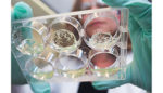
The technology could help people with diabetes or those who have had their pancreas removed due to severe pancreatitis. The team of researchers used the hydrogel material and protein combo to evaluate multiple sites for islet cell cluster implantation.
“We have engineered a material that can be used to transplant islets and promote vascularization and survival of the islets to enhance their function,” Andrés García, a professor at Georgia Institute of Technology, said in prepared remarks. “We are very excited about this because it could have immediate patient benefits if this proves successful in humans.”
Although doctors have been transplanting pancreatic islet cells into humans for years, many of the transplated cells die immediately. The cells are cut off from their blood supply and they are often killed by an immune response.
In previous testing, islet cells have been placed into the liver’s vasculature, where there is a hefty supply of blood. But there, the cells also face a tough immune environment.
The team devised a degradable polymer hydrogel material used to deliver the transplanted cells and incorporated a protein, vascular endothelial growth factor, to stimulate the growth of blood vessels into transplanted cells.
“The transplanted islets need a lot of oxygenation and a connection to the body’s circulatory system to sense the glucose levels and transport the insulin,” García said. “In addition to protecting the islets, our engineered material promotes the formation of new blood vessels to nourish the cells.”
The protein, VEGF, is tricky – use too much and it will trigger the formation of leaky blood vessels. Use too little and the vessels don’t grow quickly enough to support the transplanted islets.
The researchers used diabetic mice to compare varying locations where the transplanted cells could be placed, including the liver, the skin and sites in the abdomen.
“We were able to study the transplant sites in parallel and really look at the pros and cons of each to compare the survival rates of the cells in each area,” 1st author Jessica Weaver said. “Islet cells are very precious because we get so few from each donor. We need them all to survive to help a patient with Type I diabetes get off insulin.”
Weaver studied the animals for 100 days and saw that cell clusters transplanted with the hydrogel-protein combo formed blood vessels and engrafted into their new locations. The hydrogel material disappeared and was eventually replaced by new tissue.
The team discovered that the abdominal fat pad was the most optimal transplant location among the sites tested. An equivalent structure exists in human beings, called the omentum.
Next, the Georgia Tech researchers want to study the technique in larger animals and eventually bring it to human clinical trials. The team said it hopes stem cells could someday act as a source of islets that doesn’t require a cadaver donor and immune system suppression.

Good evening…I have 13 year old identical twin boys…one of which was diagnosed with T1D last November. I’ve had his twin,as well as the rest of the family, tested…all with negative results. I’m wandering if there is testing in identical for beta cell transplantation?