Drug Delivery Business

  • Clinical Trials
  • Research & Development
  • Drug-Device Combinations
  • FDA
  • Pharmaceuticals
  • Policy

This engineered heart ventricle helps with studying arrhythmia and cardiomyopathy

July 26, 2018 By Danielle Kirsh

engineered-heart-ventricle-vector-blog

[Luke MacQueen and Michael Rosnach/Harvard University]

While engineered heart tissues can replicate muscle contraction and electrical activity in a dish, many aspects of heart disease can only adequately be captured in 3D. In a report published online yesterday by Nature Biomedical Engineering, researchers describe a scale model of a heart ventricle, built to replicate the chamber’s architecture, physiology and contractions. Cardiac researchers at Boston Children’s Hospital think it could help them find treatments for congenital heart diseases.

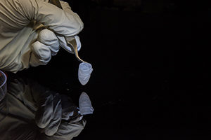
Building a 3D engineered heart ventricle

Collaborators from the Harvard School of Engineering and Applied Sciences (SEAS), the Wyss Institute for Biologically Inspired Engineering, the Harvard Stem Cell Institute and Boston Children’s engineered the ventricles by seeding rat or human heart cells onto 3D nanofiber scaffolds made of biodegradable polyester and gelatin fibers.

Guided by the scaffold, the cells aligned and assembled into beating ventricle chambers. The ventricles’ pressure, volume and contraction rate could be measured as they are in patients. In one test, for example, their beat rate increased when exposed to an adrenaline-like drug.

“The applications, from regenerative cardiovascular medicine to its use as an in vitromodel for drug discovery, are wide and varied,” said Kit Parker, PhD, of SEAS, senior author of the study, in a press release.

Studying cardiomyopathies and arrhythmias

Coauthor William Pu, MD, director of Basic and Translational Cardiovascular Research at Boston Children’s, provided the human cells used for the study. He wants to use the engineered ventricles to better understand genetic forms of heart failure and rhythm disturbances that he sees in children with congenital heart disease.

“The current step was just to develop the technology,” says Pu, who is collaborating with Parker under an NIH “Tissue Chips 2.0” grant. “The next step would be to use these ventricles to model disease.”

The final goal, of course, is to test treatments.

Read the full blog post: “A tissue engineered heart ventricle for studying rhythm disorders, cardiomyopathy” on Vector Blog.

The opinions expressed in this blog post are the author’s only and do not necessarily reflect those of DrugDeliveryBusiness.com or its employees.

Filed Under: Cardiovascular Tagged With: blog, Vector Blog

IN CASE YOU MISSED IT

  • Leo Pharma, Portal Instruments partner to develop needle-free drug delivery device
  • J&J’s Ethicon launches Vistaseal fibrin sealant
  • Senseonics launches Eversense CGM System with non-adjunctive claim
  • ‘Hubble Space Telescope’ for genomics could yield new drugs, materials
  • OncoSec $30m fundraise threatened by spat
Need Drug Delivery Business News in a minute? We Deliver!
Drug Delivery Enewsletters get you caught up on all the mission critical news you need in med tech. Sign up today.

Tweets by DrugDeliveryNow

MassDevice Medical NETWORK

MassDevice
DeviceTalks
Medical Design & Outsourcing
Medical Tubing + Extrusion

DRUG DELIVERY BUSINESS NEWS

Subscribe to Drug Delivery News
Advertise with us
About
Contact us
Privacy
Add us on FacebookDrug Delivery Business News
Follow us on Twitter@DrugDeliveryNow
Connect with us on LinkedInLinkedIn

Copyright © 2019 · WTWH Media LLC and its licensors. All rights reserved.
The material on this site may not be reproduced, distributed, transmitted, cached or otherwise used, except with the prior written permission of WTWH Media.

Advertise | Privacy Policy | RSS